
Services
China Top Image Intensifier Fluoroscopy Manufacturers?
China’s medical imaging sector is expanding, but choosing reliable Image Intensifier Fluoroscopy manufacturers requires more than comparing prices. Buyers must examine detector performance, radiation control, mechanical stability, software integration, and long-term service support. A clear evaluation method matters.
The International Atomic Energy Agency emphasizes optimization and justification in fluoroscopy-based procedures. Its radiation protection guidance encourages facilities to monitor dose, improve operator training, and use equipment suited to clinical requirements. The UNSCEAR 2020/2021 report also identifies medical exposure as the main human-made source of ionizing radiation. These findings make dose management a purchasing priority, not a marketing detail.
China offers a broad manufacturing base, from established medical device groups to specialized OEM suppliers. Leading companies may provide compact C-arms, mobile fluoroscopy systems, replacement image intensifiers, and integrated surgical imaging platforms. However, product quality can vary between similar-looking systems. It can be easy to overlook service response time, calibration support, spare-part availability, and local registration status.
The World Health Organization’s Global Patient Safety Action Plan 2021–2030 supports safer diagnostic and treatment technologies. This principle should guide supplier selection. Look for documented testing, transparent technical specifications, verified quality systems, and practical installation experience. Ask how the manufacturer handles tube replacement, image calibration, and preventive maintenance.
That question is harder than it looks.
A strong shortlist should balance clinical evidence, engineering capability, export experience, and after-sales reliability. No ranking is perfect. Hospital workloads differ, and a system that performs well in one operating room may not suit another. This guide reviews China’s top Image Intensifier Fluoroscopy manufacturers through those practical and evidence-based criteria.
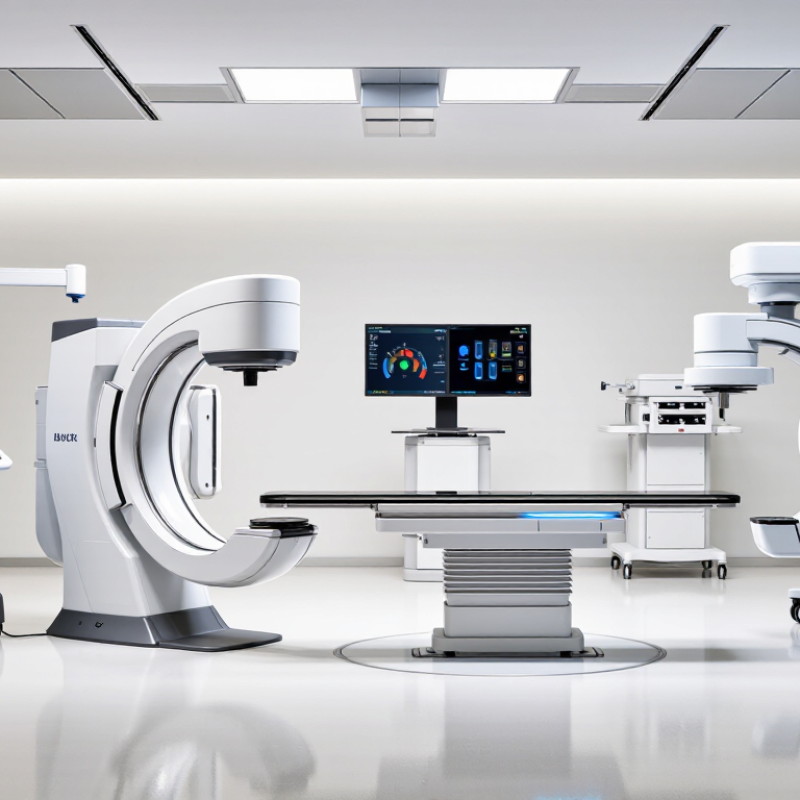
What Is an Image Intensifier Fluoroscopy System?
An image intensifier fluoroscopy system creates real-time X-ray images during movement, such as swallowing, catheter placement, or orthopedic positioning. The image intensifier first converts X-rays into light, then electrons amplify that signal before a display shows the image. Its vacuum tube, input window, electrostatic lenses, and output screen work together. The system does not produce a static photograph. It follows motion.
The U.S. Food and Drug Administration states that fluoroscopy radiation exposure depends on dose rate, exposure time, patient size, and operating mode. The FDA also identifies 5 Gy skin dose as a level requiring clinical attention. The International Atomic Energy Agency recommends pulsed fluoroscopy, last-image hold, tight collimation, and careful distance management. These controls can reduce unnecessary exposure, although results vary between procedures. The terminology is easy to oversimplify. Image quality still depends on operator judgment, not only hardware.
Tips: Check automatic brightness control, magnification modes, detector alignment, and dose-area-product readings during acceptance testing. Ask for measured performance data, not only brochure claims. A clinical engineer should verify uniformity, spatial resolution, and entrance-surface dose under realistic patient conditions. Industry surveys often report efficiency improvements, but older image intensifiers may show geometric distortion, veiling glare, or reduced contrast. That weakness deserves honest inspection. Calibration records matter. So does staff training.
China Top Image Intensifier Fluoroscopy Manufacturers? - What Is an Image Intensifier Fluoroscopy System?
| Evaluation Dimension | Typical Technical Data or Requirement | What It Means in Practice | Recommended Verification Point |
|---|---|---|---|
| System Definition | An image intensifier fluoroscopy system converts X-rays into visible light and then into an amplified electronic image for real-time viewing. | It supports dynamic imaging during procedures such as gastrointestinal studies, pain management, orthopedic surgery, and vascular examinations. | Confirm the system’s intended clinical applications and compatible X-ray generator. |
| Input Field Sizes | Common nominal field sizes include approximately 9, 6, and 4.5 inches, depending on the model and selected magnification mode. | The larger field provides broader anatomical coverage, while smaller fields improve detail by electronically magnifying the image. | Check active field diameter, number of selectable fields, and the available image area at each mode. |
| Image Conversion Chain | The main stages are input phosphor, photocathode, electrostatic focusing, anode acceleration, output phosphor, optical coupling, and camera or digital acquisition. | Each stage affects brightness, contrast, spatial resolution, image lag, and radiation efficiency. | Request the detector construction, optical coupling method, and image-acquisition interface specifications. |
| Spatial Resolution | Typical image-intensifier systems are commonly specified in line pairs per centimetre, with resolution varying by field size and operating mode. | Resolution generally improves when a smaller input field is selected, although the visible anatomy area becomes smaller. | Compare measured resolution at every field size rather than relying only on the maximum value. |
| Contrast Performance | Contrast depends on X-ray spectrum, scatter control, image-intensifier design, automatic exposure control, and image-processing settings. | Higher contrast helps distinguish anatomical structures and devices when tissue attenuation differences are small. | Ask for contrast-detail test results under clinically relevant exposure conditions. |
| Brightness Gain | Brightness gain is produced by minification gain and flux gain within the image intensifier. It allows fluoroscopy to operate with less radiation than a conventional fluorescent screen. | The system can create a usable real-time image while helping control patient and operator exposure. | Review brightness-gain data together with patient entrance dose and image-noise measurements. |
| Automatic Brightness or Dose Control | Automatic brightness control adjusts tube voltage, tube current, pulse width, or other exposure parameters to maintain image brightness. | It compensates for changes in patient thickness, anatomy, collimation, and magnification mode. | Verify response time, exposure limits, selectable dose modes, and compatibility with pulsed fluoroscopy. |
| Pulsed Fluoroscopy | Pulsed operation generates X-ray exposures at selected pulse rates instead of continuously producing radiation. | Lower pulse rates can reduce dose, while higher pulse rates provide smoother motion for fast procedures. | Check available pulse rates, pulse-width control, temporal resolution, and image lag. |
| Geometric Distortion | Image intensifiers may exhibit pincushion distortion, S-distortion, and veiling glare because of their curved input surface and electron-optical design. | Distortion can affect the apparent position, shape, or size of anatomical structures and instruments. | Request distortion specifications, calibration procedures, and correction capability in the digital image chain. |
| Image Lag | Image lag is the persistence of a previous image after the X-ray exposure changes or stops. | Low lag is important for moving anatomy, guidewire placement, and rapid instrument manipulation. | Evaluate lag using dynamic test objects and clinically representative pulse rates. |
| Digital Image Output | Common digital interfaces may include video output, Ethernet-based transfer, DICOM connectivity, and USB or service interfaces, depending on the system design. | Digital output enables recording, post-processing, reporting, archiving, and integration with hospital information systems. | Confirm image format, bit depth, frame rate, DICOM functions, and export workflow. |
| Radiation Safety | Important controls include collimation, filtration, pulsed exposure, last-image hold, dose-rate display, exposure-time monitoring, and protective shielding. | These functions help reduce unnecessary radiation exposure for patients and clinical staff. | Check dose-area-product or cumulative-dose display, warning thresholds, and compliance documentation. |
| Mechanical Integration | The image intensifier may be mounted on a C-arm, U-arm, table system, or other fluoroscopic support structure. | Travel range, angulation, clearance, and positioning accuracy directly influence procedural access. | Verify focal-spot-to-input distance, source-to-image distance, rotation range, collision protection, and load ratings. |
| Quality and Compliance | A qualified supplier should provide documented quality management, electrical safety testing, electromagnetic compatibility testing, and applicable medical-device regulatory documentation. | Traceable documentation supports procurement, installation, acceptance testing, maintenance, and regulatory audits. | Request certificates, test reports, risk-management records, user manuals, and acceptance-test procedures. |
| Service and Lifecycle Support | Key factors include spare-part availability, calibration support, preventive-maintenance intervals, technical training, warranty terms, and response time. | Strong lifecycle support helps maintain image quality, system uptime, and consistent radiation performance. | Evaluate service coverage, replacement-part lead time, remote diagnostics, and documented preventive-maintenance procedures. |
Note: Technical specifications vary by system configuration, image-intensifier size, X-ray generator, camera chain, and clinical application. Final selection should be based on verified test data and applicable local medical-device requirements.
Related Posts
-
Top 10 Fluoro Image Intensifier Manufacturers in 2026
-
How to Enhance Night Vision Using Mcp Image Intensifiers
-

Top Digital Image Intensifier Tips for Global Buyers?
-
What Is Image Intensifier Night Vision and How Does It Work?
-
2026 Best UV Image Intensifiers for Global Buyers
-

Innovative Solutions for Sourcing Fluoro Image Intensifiers Globally