Top 10 Fluoro Image Intensifier Manufacturers in 2026
Fluoro Image Intensifier technology remains important in mobile C-arms, gastrointestinal studies, vascular imaging, and older fluoroscopic systems. Although flat-panel detectors dominate new installations, image intensifiers still serve thousands of hospitals, clinics, and repair departments worldwide. Their circular output window, visible geometric distortion, and dependable low-dose performance remain familiar to many radiology professionals.
This 2026 overview examines ten leading Fluoro Image Intensifier manufacturers. The selection considers product reliability, image quality, input-field options, replacement support, technical documentation, and long-term market presence. It also considers practical details, such as compatibility with existing C-arms, connector design, housing dimensions, and service availability. These factors matter when a replacement unit must fit an aging imaging system without causing costly downtime.
Dr. John W. Coltman, recognized as a pioneer in X-ray image intensifier development, described the central engineering goal as “making the X-ray image brighter without losing useful detail.” That principle still matters.
But rankings are not absolute.
A manufacturer may lead in production scale yet offer limited regional support. Another may provide excellent custom engineering but fewer standard models. Performance can also change after installation, especially when calibration, shielding, and maintenance are inconsistent. This is where our evaluation remains imperfect. Public specifications rarely reveal every field failure, service delay, or compatibility issue.
Readers should therefore use this list as a research starting point, not a purchasing verdict. The strongest choice depends on clinical workload, system age, budget, regulatory requirements, and the availability of qualified service engineers.
What Fluoro Image Intensifiers Are and How They Work
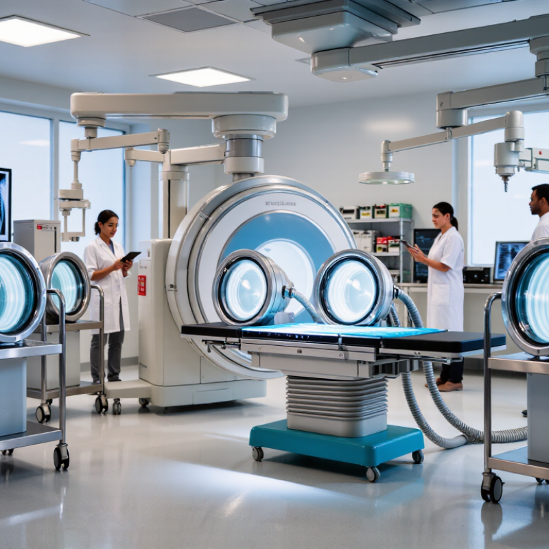
A fluoro image intensifier converts low-level X-rays into a brighter image for real-time medical imaging.
It sits inside the fluoroscopy system, between the patient and the image processing chain. The input window receives X-rays, while an input phosphor changes them into visible light. A photocathode then converts that light into electrons. These electrons are focused and accelerated through an electric field.
The electrons strike an output phosphor and create a much brighter image.
A camera or digital sensor captures this image for display. In practice, the process is remarkably fast. It helps clinicians observe moving anatomy during procedures, including changes that would disappear in a still radiograph. The device may also offer different field sizes, allowing close inspection or broader anatomical coverage.
Real performance is not perfectly clean.
Images can show pincushion distortion, veiling glare, or reduced sharpness near the edges. The input screen may also lose efficiency after years of use. Small defects matter.
Technicians should check brightness, contrast, geometric accuracy, and image lag during routine quality control.
A poorly calibrated system can make anatomy appear less reliable than it should. The technology remains useful, but its limitations deserve more attention than they often receive.
Key Factors for Evaluating Manufacturers in 2026
For a 2026 top-ten list, manufacturer evaluation should begin with measurable clinical performance, not brochure language.
Compare spatial resolution, contrast detail, distortion, lag, and image stability under continuous fluoroscopy. Ask for acceptance-test records from installed systems. A polished demo is not enough.
Dose management deserves equal weight.
UNSCEAR reports that medical exposure accounts for about 98% of artificial ionizing-radiation exposure to the global population. The U.S. FDA notes that high-level fluoroscopy modes can reach 20 cGy per minute at the skin. Therefore, assess pulsed operation, automatic dose-rate control, last-image hold, and audible dose alerts. Require testing against applicable IEC 60601 standards, with clear service documentation.
Reliability is harder to judge. It still matters most.
Request field failure rates, mean time to repair, spare-part availability, detector replacement terms, and software-support duration. ISO 13485 certification supports process control, but it does not guarantee excellent clinical outcomes. Independent acceptance testing remains necessary. I would also compare five-year ownership cost, including calibration, cooling systems, and emergency service. MarketsandMarkets’ 2024 fluoroscopy and C-arms analysis indicates continued equipment-market expansion, yet growth does not prove equal quality. A lower purchase price may hide downtime. This is where many rankings become too confident.
Innovations Shaping Fluoroscopy Imaging Technology
Top 10 Fluoro Image Intensifier Manufacturers in 2026
Fluoroscopy imaging is changing through smarter image intensifiers, improved dose control, and faster clinical feedback. Modern systems use cesium iodide input screens to capture clearer images with less radiation. Better optical coupling also helps preserve detail across the viewing field.
Pulsed fluoroscopy now works with adaptive exposure settings. The system can adjust frame rates when motion slows or stops. That matters during vascular procedures and orthopedic positioning. New processing algorithms reduce noise without making guidewires disappear. Useful progress. However, excessive processing may create artificial edges and mislead inexperienced users.
From a clinical engineering perspective, reliability remains as important as resolution. Manufacturers are improving thermal stability, mechanical protection, and calibration software. Service teams can detect output changes before image quality visibly declines. Independent testing should still verify dose, contrast, lag, and geometric distortion. Marketing claims are not enough.
A strong 2026 evaluation should include workflow tests, not only laboratory charts. Operators need responsive controls, readable displays, and consistent images at different angles. Artificial intelligence may support image enhancement, but it cannot replace trained judgment. I would also question systems that hide technical limitations behind impressive demonstrations. No device is perfect. Real operating rooms are messier than brochures.
Top 10 Fluoro Image Intensifier Manufacturers in 2026 – Innovations Shaping Fluoroscopy Imaging Technology
Representative pulsed-fluoroscopy operating points used to illustrate how lower pulse rates can reduce exposure while preserving clinically useful motion imaging. The exposure index is normalized to 30 pulses/s.
Note: Actual patient dose depends on equipment settings, anatomy, filtration, automatic exposure control, field size, and procedure type.
How to Choose the Right Manufacturer for Your Application
Choosing among the Top 10 Fluoro Image Intensifier Manufacturers in 2026 requires more than comparing prices.
Start with the clinical task. A mobile C-arm for orthopedic work needs compact geometry, fast positioning, and stable output during repeated exposures. A vascular suite demands finer contrast, reliable cooling, and precise dose management. Ask for measured performance at your operating dose, not only brochure resolution.
Grand View Research estimated the global fluoroscopy systems market at about
USD 1.9 billion in 2023, with projected growth near
5.5% annually through 2030. Market definitions vary, so treat this figure as directional evidence rather than a purchasing formula. The IAEA’s Radiation Protection and Safety guidance stresses that patient and staff doses can differ greatly between procedures. Therefore, compare automatic exposure control, dose reporting, shielding compatibility, and service response times. A low purchase price may become expensive when calibration delays interrupt a full procedure list.
Request acceptance-test records, thermal limits, image-retention data, and a clear warranty scope.
Confirm compatibility with your generator, detector housing, operating table, and hospital network. IEC 60601 compliance matters, but certification alone does not prove clinical suitability. Visit a reference site if possible. Watch the image during motion, not only on a still test screen. Some evaluation methods remain too optimistic. I have seen specifications hide practical weaknesses, especially after long fluoroscopy sessions. A realistic trial should include your staff, your workflow, and difficult anatomy.
Reliability is experienced in the room.